

 You Are Here:
Fully Automated Exosome Separation Using Multistage Filtration and Performance Evaluation
 Last updated: 2023-04-06
 Views: 2019
A fully automated multistage-filtration instrument for separating exosomes was developed and its performance evaluated by extracting exosomes derived from human autologous adipose stem cells.Methods:Define the instrument’s separation process, design its structure, and complete hardware and software development;obtain autologous adipose tissue from patients, digest it to isolate adipose stem cells, and culture and passage the cells. Collect culture supernatant from third-passage adipose stem cells and perform preliminary centrifugation. Add the supernatant to the first-stage reservoir and filter it with the instrument, then centrifuge the third-stage filtrate to extract exosomes. Usetransmission electron microscopy(Transmission Electron Microscope,TEM) to observe exosome morphology and perform nanoparticle tracking analysis (Nanoparticle Tracking Analysis,NTA),to analyze particle size and concentration, and use Western blotting (Western Blot) to detect expression of proteins specific to adipose-stem-cell-derived exosomes. Expression markers includeCD63、CD81、TSG101, while also detectingGM130to determine the degree of vesicle contamination in the tested sample.Results:Completed thedevelopment of the fully automated multistage-filtration exosome-separation instrument. Adipose-stem-cell-derived exosomes obtained after filtration, concentration and centrifugation appeared round and cup-shaped under transmission electron microscopy. Nanoparticle tracking analysis showed that most exosomes were within50nm-200nm; Western blotting showed that the extracted exosomes expressedCD63、CD81、TSG101, with an exosome yield reaching75%, and vesicle contamination within the normal range.Conclusion:The fully automated multistage-filtration instrument separates exosomes rapidly and efficiently, meets the requirements for clinical application, and provides strong support for clinical exosome-based disease treatment.
Keywords:exosomes; adipose stem cells; filtration; concentration; extraction
Multistage Filtration Based Fully Automated Exosomes Separation Instrument and Its Performance Evaluation
Zeng Yangtian1 Zheng Yi2 Liu Kai2* Li Hong1*
1(Hangzhou Baiqiao Medical Technology Co.,Ltd., Hangzhou 310018)
2 (Shanghai Ninth People's Hospital, Shanghai Jiao Tong University School of Medicine, Shanghai 200011)
3 (EVERFINE Test and Calibration Technology Co., Ltd, Hangzhou, Hangzhou 310053)
Abstract: Objective: to develop a fully automated multistage filtration instrument for the separation of exosomes and to evaluate the performance by extracting human autologous adipose-derived stem cells(ADSCs) exosomes. Methods: To determine the instrument separation process, design the instrument structure and complete the instrument development of both hardware and software; to obtain and digest the autologous fat from patients, obtain and subculture the adipose-derived stem cells, collect the supernatant of the third generation adipose-derived stem cell culture and make initial centrifugation, add the supernatant to the first reservoir then filter through the instrument, centrifuge the obtained third stage filtrate and extract exosomes. The morphology of the exosomes was observed by Transmission Electron Microscope (TEM). Nanoparticle Tracking Analysis (NTA) was performed to analyze particle size and concentration. Western Blot (WB)was used to detect the specific protein expression of the adipose-derived stem cells exosomes, which including CD63, CD81, TSG101 and GM130.And the Western Blot Method also was used to detect the contamination extent of vesicles in the assay samples. Results: The development of a fully automated multistage filtration and separation instrument for exosomes was completed. The adipose-derived stem cell exosomes, which extracted by the instrument after filtration and concentration and centrifugation, were observed in a circular-cup shape by TEM. Most of the exosomes obtained by NTA were in the range of 50 nm-200 nm. WB showed that the extracted exosomes contained the expression of CD63, CD81, TSG101 and CD81. Exosomes were obtained up to 75% and vesicle contamination was within the normal range. Conclusion: The fully automated multistage filtration and separation of exosomes instrument can achieve efficient and rapid separation of exosomes, which meets the requirements of clinical application and provides a strong support for clinical exosomes in disease treatment.
Keywords: Exosomes; Adipose-Derived Stem Cells; Filtration; Concentration; Extraction
Introduction
Exosomes (exosome) are extracellular vesicles with diameters of30~150nmextracellular vesicles[1-3]; studies[4-8]show that exosomes are widely present in body fluids such as urine, pleural and peritoneal effusions, milk and saliva. Many types of cells can secrete exosomes, includingMSCs, blood cells, endothelial cells, neurons and tumor cells. Exosomes carry extensive intracellular and extracellular biological information, act as mediators of cell communication and extracellular-matrix remodeling, and are closely linked to human health and disease[9-10],through aerosol inhalationMSC-EVsas a route of administration has major significance for treating acute respiratory distress syndrome[11]; Zhu Hongyan’s[12]research group showed that:CBSA/siS100A4can significantly inhibit the growth of malignant breast-cancer cells; Ma Taotao’s[13]team revealed that exosome-mediated cross-talk between renal tubular epithelial cells and macrophages plays an important role in treating diabetic nephropathy; Zhang Songying’s[14]team found that regulating decidualNKcell (maternal) metabolism thereby improves decidualNKcell secretion of factors required for placental development.As the value of exosomes in clinical diagnosis, disease treatment and tissue regeneration has become increasingly evident in recent years, researchers worldwide have studied their distinctive properties and structure in ever greater depth.
Traditional exosome-separation methods include ultracentrifugation, filtration, immunomagnetic beads and microfluidic chips[15]. Ultracentrifugation is difficult to adopt widely because its equipment is expensive. Immunomagnetic beads introduce foreign components that limit clinical use. Microfluidic chips achieve high separation purity but produce very small yields and are difficult to scale. Although filtration is mature, it is currently performed manually, making it labor-intensive, time-consuming and prone to contamination[16-17]。
This study improves the filtration method, optimizes filtration pore sizes and stages, and develops intelligent control of the entire process to meet clinical requirements for efficient, rapid and safe use. The instrument’s exosome-preparation performance was then evaluated experimentally.
1 Methods
1.1 Instrument Design
1.1.1 Overall Instrument Configuration
The instrument consists of two reservoirs, one multistage filtration unit, a motor drive, a plunger pump, two pressure sensors, a motor, a linear-motion mechanism, a touchscreen and other components (Figure1). The entire system is controlled byPLCand a data-acquisition module. Pressure-sensor readings are displayed on the touchscreen in real time, and the touchscreen provides the human–machine interface. The instrument offers automatic and manual modes, which can be selected for the operating scenario.
1.1.2 Instrument Workflow
The process for concentrating a target liquid with the instrument is shown in Figure2: liquid is added to Reservoir 1 and the power is switched on. The controller operates the motor drive, which starts and stops the motor. When the motor runs, the linear-motion mechanism drives the plunger pump. A suction tube draws liquid from Reservoir 1 and sends it through the piping to the multistage filtration unit. Pressure sensors are installed at the unit’s inlet and outlet to measure in real timethe difference between pressure-sensor readings in the first- and second-stage pressure-stabilizing chambers. The plunger pump’s on/off timing is adjusted according to that difference, maintaining a constant differential pressure across the multistage filtration unit. This drives liquid through the multistage filtration unit, automatically filtering particles by diameter. At the end of a run, particles smaller than the pore size of the unit’s lowest membrane and the waste liquid are discharged through the terminal line into Reservoir 2. Researchers can collect the desired product from the appropriate stage of the filtration unit.
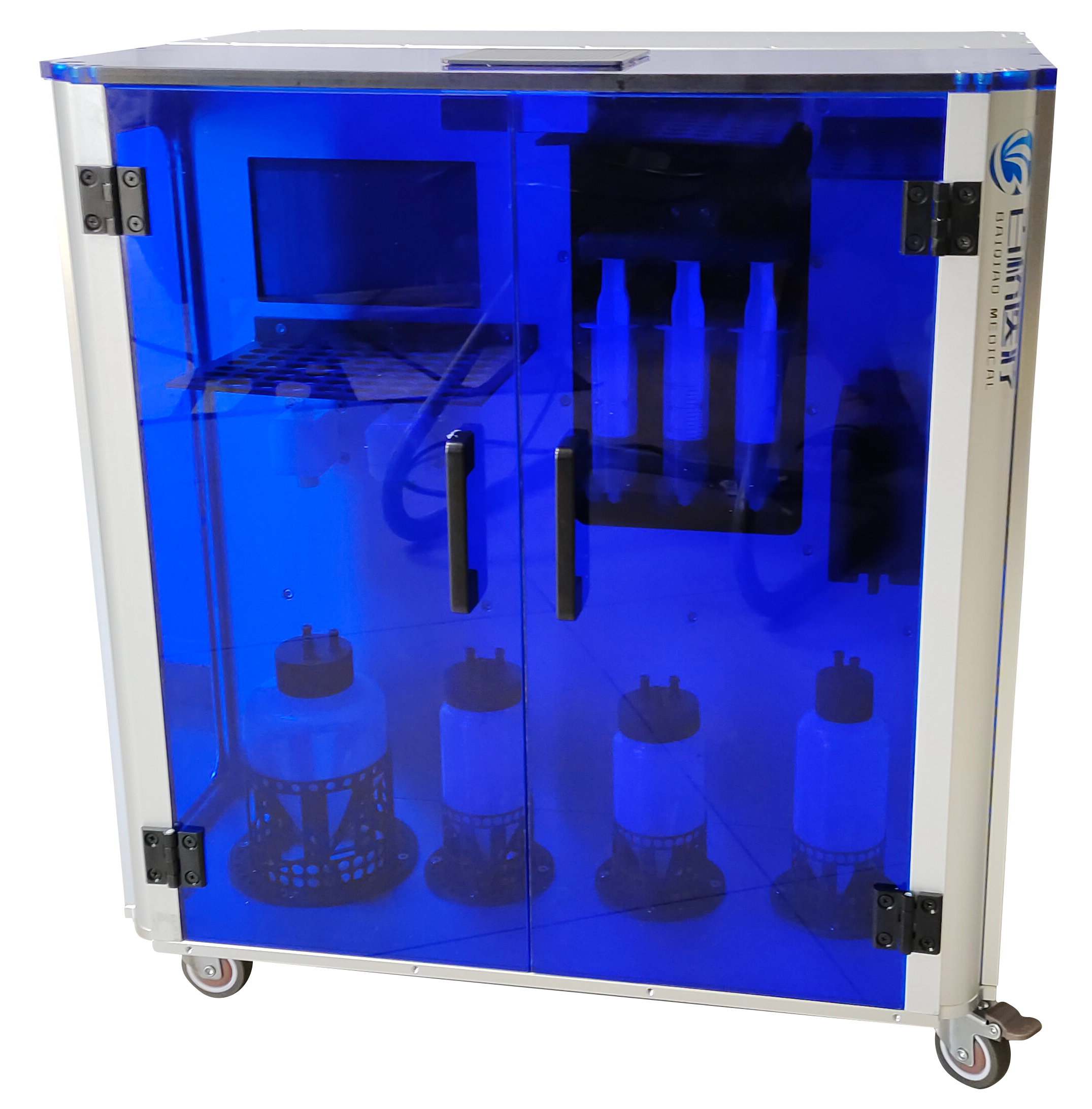
Figure1 Fully Automated ExosomeIsolation and ConcentrationInstrument
Fig.1 Automatic Exosome Filtration Instrument
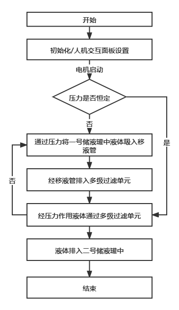
Figure2 Instrument Workflow Diagram
Fig.2 Instrument work flow chart
1.2 Instrument Performance Evaluation
1.2.1 Experimental Materials
1.2.1.1 Main Experimental Reagents
adipose stem-cell culture supplement (UltraGROTM,AventaCell BioMedical, USA);PBS(Gibco, USA);0.25%trypsin (Gibco, USA); fetal bovine serum (Gibco, USA); triple-antibiotic solution (Gibco,USA);α-MEM, low-glucose and high-glucoseDMEM(Gibco, USA); collagenase (Collagenase NB4,Serva, Germany); BCAprotein assay kit (Beyotime, China).
1.2.1.2Main Experimental Instruments
disposable sterile10 cmculture dishes (BD, USA);100 kDaultrafiltration tubes (Milipore, USA);clean bench (Suzhou Purification Equipment Co., Ltd., China); microplate reader (Thermo Fisher Scientific, USA);precision electronic balance (Longcheng Instrument Equipment Co., Ltd., China); flow cytometer (Beckman,USA);temperature-adjustable benchtop centrifuge(Thermo Fisher Scientific, USA);hemocytometer (Shanghai Qiujing Biochemical Reagent Instrument Co., Ltd., China);temperature-adjustable shaker (Taicang Huamei Biochemical Instrument Factory, China);constant-temperature water bath (Shanghai Hetian Scientific Instruments Co., Ltd., China).
1.2.2 Experimental Methods
1.2.2.1Isolation and Culture of Adipose Stem Cells
The adipose tissue in this study came from the abdomen or inner thigh and was tissue discarded after patients underwent liposuction. All patients signed informed-consent forms agreeing to donate the discarded tissue for medical research. The study protocol was approved by the Ethics Committee of Shanghai Ninth People’s Hospital, Shanghai Jiao Tong University School of Medicine.
After the donated adipose tissue was obtained, it was digested with collagenase and centrifuged to isolate primary adipose stem cells. The cells were seeded inα-MEMculture medium containing10cmculture dishes and cultured at5%CO2,37°C. When the cells approached80%-90%confluence, a solution containingEDTAof0.25%Passage the cells by trypsin digestion. Use passage3adipose-derived stem cells for exosome extraction.
1.2.2.2Extraction of adipose-derived stem cell exosomes
Transfer the collected passage3adipose-derived stem cell supernatant into the inlet reservoir of the exosome extractor and filter it with the instrument. Add the collected filtrate to a100kDaofmiliporeultrafiltration tube to extract exosomes by ultrafiltration. Centrifuge at3000g,15-20min,4℃ until nearly all supernatant in the inner ultrafiltration tube has passed through, then usePBSto wash once and centrifuge until only200-500μLof exosome-containingPBS. After centrifugation, aspirate the exosome-containingPBSand store at4℃ in a refrigerator.
1.2.2.3Identification of adipose-derived stem cell exosomes
With current separation methods, it is difficult to obtain exceptionally pure, homogeneous exosomes. The exosomes obtained by centrifuging the instrument-concentrated sample therefore need to be identified. This study used three identification methods in accordance with standards established by the International Society for Extracellular Vesicles to ensure accuracy.① Transmission electron microscopy (TEM) to observe the morphology of pretreated exosomes[18];② Size-based nanoparticle tracking analysis (NTA): a light beam illuminates particles in the exosome sample, and each particle's trajectory is recorded from its scattered light and Brownian motion. The mean velocity and diffusion coefficient can then be determined and used in mathematical calculations to obtain the exosome concentration and size distribution[19];③ Western blotting (Western Blot) to detect exosome-specific surface proteinsmarker [20]。
1.2.3 Primary observation endpoints
①transmission electron microscopyObserve the morphology of adipose-derived stem cell exosomes;②nanoparticle tracking analysis,analysis to determine exosome particle size and concentration;③ Western blot identification of proteins expressed by adipose-derived stem cell exosomes.
2 Results
2.1 Adipose-derived stem cell morphology
Figure3(a) shows washed and purified adipose tissue; Figure3(b)shows third-passage human adipose-derived stem cells with a long spindle-shaped morphology under an inverted microscope.
2.2 Transmission electron microscopy identification of exosomes
As shown in Figure4(a),under transmission electron microscopy, adipose-derived stem cell exosomes measured30nm-200nmand exhibited the typical round, cup-shaped exosome morphology.
2.3 Western blot identification of exosome markers
Under the International Society for Extracellular Vesicles standard[21], a vesicle can be called an exosome only if it contains an endosomal or exosomal plasma-membrane protein and a cytosolic protein associated with exosome secretion, while excluding a non-exosomal structural protein or broadly distributed cytosolic protein. As shown in Figure4(b), Western blotting detected expression of the exosomal plasma-membrane proteinCD63、CD81and the secretion-associatedESCRTcomplex proteinTSG101, while the Golgi proteinGM130was scarcely expressed, demonstrating that the isolated product contained exosomes of relatively high purity.
2.4 Nanoparticle tracking analysis of exosomes
As shown in Figure5shows that nanoparticle tracking analysis found the particle-size distribution to be concentrated between30nm-200nm; the particle concentration and size were consistent with exosome characteristics[22]。
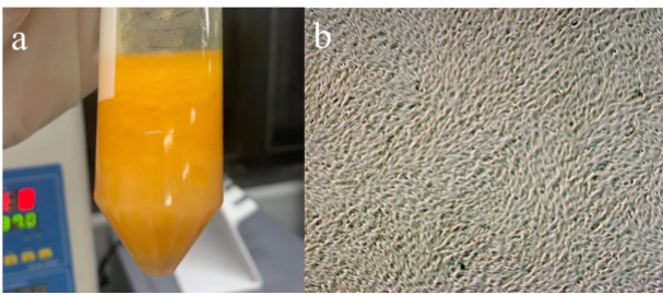
Figure3 Experimental materials. (a) Adipose tissue used in the experiment; (b) Morphology of human adipose-derived stem cells
Fig.3 Experimental materials.(a)Fat used in experiments;(b)Morphology of human adipose stem cells
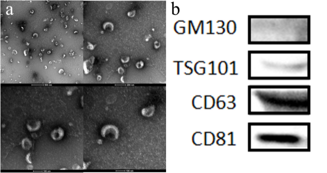
Figure4 Exosome morphology and specific-protein identification. (a) Exosome morphology shown by transmission electron microscopy; (b) Western blot detection of exosome protein expression
Fig.4 Exosome morphology and specific protein identification.(a)Morphological characteristics of exosomes displayed by transmission electron microscopy;(b)Western blotting to detect exosome protein expression
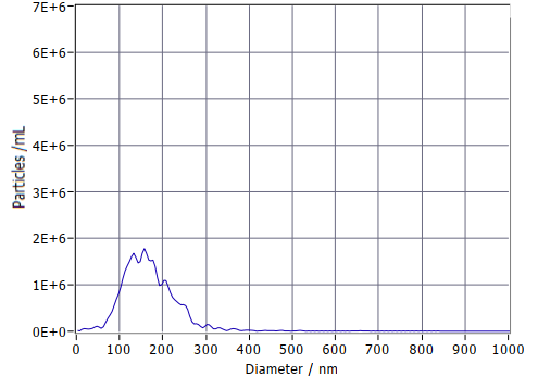
Figure5 Exosome particle-size distribution measured by NTA
Fig.5 NTA detection of exosome particle size distribution
3 Discussion
The performance evaluation of the fully automated multistage-filtration exosome separator used autologous adipose-derived stem cells as the raw material. The clinical significance of extracting exosomes from autologous adipose-derived stem cells is that autologous adipose tissue is abundant and easy to obtain, causes no rejection, and is convenient to use. In addition, thefully automated multistage-filtration exosome separator uses a non-contact design with intelligent closed-loop control, effectively reducing labor costs and producing high-purity, contamination-free exosomes that meet clinical requirements.The experimental evaluation showed that after concentrating and centrifugally extracting adipose-derived stem cell exosomes, all three standard identification methods confirmed that the extracted microparticles were exosomes and that contamination was within the normal range. This demonstrates that the fully automated exosome filtration instrument can filter and concentrate exosome-containing supernatant and isolate intact exosomes from the concentrate. The processing time required is far shorter than with traditional extraction methods and meets clinical application requirements.
The completed instrument can precisely separate exosome subpopulations by particle size (30-150nm), providing a valuable tool for studying the relationship between the structure and function of exosome subpopulations and a fast, efficient, labor-saving method for clinical treatment. The instrument still has some limitations: some exosomes adhere to the filter membrane, causing product loss and reduced activity. Future research will further improve the exosome extractor by adopting new filtration methods such as tangential flow to address this limitation, reduce concentrate waste, and increase exosome yield.
4 Conclusion
The fully automated multistage-filtration instrument separates exosomes rapidly and efficiently, meets the requirements for clinical application, and provides strong support for clinical exosome-based disease treatment.
References
[1] Raposo G,Stoorvogel W. Extracellular vesicles: exosomes, microvesicles, and friends.[J]. The Journal of cell biology,2013,200(4).
[2] Lee Y,El A S,Wood MJA. Exosomes and microvesicles: extracellular vesicles for genetic information transfer and gene therapy.[J]. Human molecular genetics,2012,21(R1).
[3] Marqués-García F,Isidoro-García M. Protocols for Exosome Isolation and RNA Profiling.[J]. Methods in molecular biology (Clifton, N.J.),2016,1434.
[4] Li P,Kaslan M,Lee SH,et al. Progress in exosome isolation tech-niques[J]. Theranostics,2017,7( 3) : 789-804.
[5] Clark DJ,Fondrie WE,Liao Z,et al. Redefining the breast cancer exosome proteome by tandem mass tag quantitative proteomics and m*riate cluster analysis[J]. Anal Chem,2015,87(20) : 10462-10469.
[6] Yu B,Zhang X,Li X. Exosomes derived from mesenchymal stem cell[J]. Int J Mol Sci,2014,15(3) : 4142-4157.
[7] Ye Dongman,Yu Tao. Extraction and Identification of Tumor Exosomes[J]. Modern Oncology,2018,26(20),3341-3343.
[8] Chan BD,Wong WY,Lee MM,et al. Exosomes in inflammation and inflammatory disease[J]. Proteomics,2019,13: e1800149.
[9] MATHIEU M, MARTIN-JAULAR L, LAVIEU G, et al. Specificities of secretion and uptake of exosomes and other extracellular vesicles for cell-to-cell communication [J]. Nat Cell Biol, 2019, 21(1): 9-17.
[10] SHU S, YANG Y, ALLEN C L, et al. Purity and yield of melanoma exosomes are dependent on isolation method [J]. J Extracell Vesicles, 2020, 9(1): 1692401.
[11] Shi MM, Yang QY, Monsel A, et al. Preclinical efficacy and clinical safety of clinical-grade nebulized allogenic adipose mesenchymal stromal cells-derived extracellular vesicles. J Extracell Vesicles. 2021;10(10):e12134.
[12] Zhao L, Gu C, Gan Y, Shao L, Chen H, Zhu H. Exosome-mediated siRNA delivery to suppress postoperative breast cancer metastasis. J Control Release. 2020;318:1-15.
[13] Jiang WJ, Xu CT, Du CL, et al. Tubular epithelial cell-to-macrophage communication forms a negative feedback loop via extracellular vesicle transfer to promote renal inflammation and apoptosis in diabetic nephropathy. Theranostics. 2022;12(1):324-339.
[14] Jiang L, Fei H, Jin X, et al. Extracellular Vesicle-Mediated Secretion of HLA-E by Trophoblasts Maintains Pregnancy by Regulating the Metabolism of Decidual NK Cells. Int J Biol Sci. 2021;17(15):4377-4395.
[15] WEN S W, LIMA L G, LOBB R J, et al. Breast cancer-derived exosomes reflect the cell-of-origin phenotype [J]. Proteomics, 2019, 19(8): e1800180.
[16] Yang Dongbin,Zhang Weihong,Zhang Huanyun,Zhang Fengqiu,Chen Lanmei,Ma Lixia,Larcher Leon M,Chen Suxiang,Liu Nan,Zhao Qingxia,Tran Phuong H L,Chen Changying,Veedu Rakesh N,Wang Tao. Progress, opportunity, and perspective on exosome isolation - efforts for efficient exosome-based theranostics.[J]. Theranostics,2020,10(8).
[17] Bai Jun,Ren Jun,Wang Jun,Zhang Hongmei,Yang Jingyue,Zhang Liwang.Experimental Study on Exosome Extraction by Centrifugal Filtration[J].Journal of the Fourth Military Medical University,2004(10):890-892.
[18] Lässer C,Eldh M,Lötvall J. Isolation and characterization of RNA-containing exosomes.[J]. Journal of visualized experiments : JoVE,2012(59).
[19] Xiao-Xia Yang,Chao Sun,Lei Wang,Xiu-Li Guo. New insight into isolation, identification techniques and medical applications of exosomes[J]. Journal of Controlled Release,2019,308(C).
[20] Théry C,Amigorena S,Raposo G,Clayton A. Isolation and characterization of exosomes from cell culture supernatants and biological fluids.[J].Current protocols in cell biology,2006,Chapter 3(1).
[21] Théry C, Witwer KW, Aikawa E, et al. Minimal information for studies of extracellular vesicles 2018 (MISEV2018): a position statement of the International Society for Extracellular Vesicles and update of the MISEV2014 guidelines. J Extracell Vesicles. 2018;7(1):1535750. Published 2018 Nov 23. doi:10.1080/20013078.2018.1535750
[22] Wang Shuai,Li Chaoran,Zhang Qianru,Huang Guilin.Extraction and Identification of Exosomes Derived from Human Urine-Derived Stem Cells[J].Chinese Journal of Tissue Engineering Research,2022,26(13):2056-2061.