

 현재 위치:
다단 여과 기반 전자동 엑소좀 분리 장비 및 성능 평가
 업데이트 날짜: 2023-04-06
 조회수: 2019
전자동 다단 여과 엑소좀 분리 장비를 개발하고 사람 자가 지방줄기세포 유래 엑소좀을 추출하여 성능을 평가했습니다.방법:장비 분리 공정을 확정하고 구조를 설계하여 하드웨어·소프트웨어 개발을 완료했습니다.환자에게서 자가 지방을 채취·소화하여 지방줄기세포를 얻고 배양·계대했습니다. 3세대 배양 상등액을 수집해 1차 원심분리하고 상등액을 1단계 저장 탱크에 넣어 장비로 여과했습니다. 3단계 여과액을 원심분리해 엑소좀을 추출했습니다.투과전자현미경(Transmission Electron Microscope,TEM)으로 엑소좀 형태를 관찰하고 나노입자 추적 분석(Nanoparticle Tracking Analysis,NTA),으로 입자 크기와 농도를 분석했으며 웨스턴 블롯(Western Blot)으로 지방줄기세포 유래 엑소좀의 특이 단백질 발현을 검사했습니다. 발현 지표는CD63、CD81、TSG101이며 동시에GM130을 검사하여 검체 내 소포 오염 정도를 판단했습니다.결과:완료한전자동 다단 여과 엑소좀 분리 장비로 여과·농축하고 원심분리해 추출한 지방줄기세포 유래 엑소좀은 투과전자현미경에서 원형 컵 모양이었으며 나노입자 추적 분석 결과 대부분50nm-200nm사이였습니다. 웨스턴 블롯 결과 추출 엑소좀에서CD63、CD81、TSG101발현이 확인되었고 엑소좀 회수량은75%에 도달했으며 소포 오염은 정상 범위였습니다.결론:전자동 다단 여과 장비는 엑소좀을 빠르고 효율적으로 분리하여 임상 적용 요건을 충족하고 엑소좀 기반 질환 치료를 지원합니다.
키워드:엑소좀; 지방줄기세포; 여과; 농축; 추출
Multistage Filtration Based Fully Automated Exosomes Separation Instrument and Its Performance Evaluation
Zeng Yangtian1 Zheng Yi2 Liu Kai2* Li Hong1*
1(Hangzhou Baiqiao Medical Technology Co.,Ltd., Hangzhou 310018)
2 (Shanghai Ninth People's Hospital, Shanghai Jiao Tong University School of Medicine, Shanghai 200011)
3 (EVERFINE Test and Calibration Technology Co., Ltd, Hangzhou, Hangzhou 310053)
Abstract: Objective: to develop a fully automated multistage filtration instrument for the separation of exosomes and to evaluate the performance by extracting human autologous adipose-derived stem cells(ADSCs) exosomes. Methods: To determine the instrument separation process, design the instrument structure and complete the instrument development of both hardware and software; to obtain and digest the autologous fat from patients, obtain and subculture the adipose-derived stem cells, collect the supernatant of the third generation adipose-derived stem cell culture and make initial centrifugation, add the supernatant to the first reservoir then filter through the instrument, centrifuge the obtained third stage filtrate and extract exosomes. The morphology of the exosomes was observed by Transmission Electron Microscope (TEM). Nanoparticle Tracking Analysis (NTA) was performed to analyze particle size and concentration. Western Blot (WB)was used to detect the specific protein expression of the adipose-derived stem cells exosomes, which including CD63, CD81, TSG101 and GM130.And the Western Blot Method also was used to detect the contamination extent of vesicles in the assay samples. Results: The development of a fully automated multistage filtration and separation instrument for exosomes was completed. The adipose-derived stem cell exosomes, which extracted by the instrument after filtration and concentration and centrifugation, were observed in a circular-cup shape by TEM. Most of the exosomes obtained by NTA were in the range of 50 nm-200 nm. WB showed that the extracted exosomes contained the expression of CD63, CD81, TSG101 and CD81. Exosomes were obtained up to 75% and vesicle contamination was within the normal range. Conclusion: The fully automated multistage filtration and separation of exosomes instrument can achieve efficient and rapid separation of exosomes, which meets the requirements of clinical application and provides a strong support for clinical exosomes in disease treatment.
Keywords: Exosomes; Adipose-Derived Stem Cells; Filtration; Concentration; Extraction
서론
엑소좀(exosome)은 직경30~150nm의 세포외소포입니다[1-3]. 연구[4-8]에 따르면 엑소좀은 소변, 흉·복강 삼출액, 모유, 타액 등 체액에 널리 존재하며MSCs혈액세포, 내피세포, 신경세포, 종양세포 등 많은 세포가 분비합니다. 엑소좀은 세포 안팎의 생물학적 정보를 운반하고 세포 소통과 세포외기질 재형성의 매개체로서 건강·질환과 밀접합니다.[9-10],분무 흡입을 통한MSC-EVs투여 경로는 급성호흡곤란증후군 치료에 큰 의미가 있습니다.[11]; Zhu Hongyan[12]연구팀은 다음을 밝혔습니다:CBSA/siS100A4은 악성 유방암 세포 성장을 현저히 억제합니다. Ma Taotao[13]연구팀은 엑소좀 매개 신세뇨관 상피세포-대식세포 상호소통이 당뇨병성 신증 치료에 중요함을 밝혔습니다. Zhang Songying[14]교수팀은 탈락막NK세포(모체 유래)의 대사 수준을 조절하여 탈락막NK세포가 태반 발달에 필요한 인자를 분비하는 수준을 개선함을 발견했습니다.최근 엑소좀이 임상 진단, 질환 치료, 조직 재생에서 중요해지며 전 세계 연구자가 특성과 구조를 더 깊이 연구하고 있습니다.
기존 엑소좀 분리법에는 초원심분리, 여과, 면역자성 비드, 미세유체 칩 등이 있습니다.[15]초원심분리는 장비가 비싸 보급이 어렵고, 면역자성 비드는 외래 성분 혼입으로 임상 적용이 어렵습니다. 미세유체 칩은 순도가 높지만 추출량이 적고, 여과법은 성숙했으나 수작업이라 힘과 시간이 들고 오염되기 쉽습니다.[16-17]。
본 연구는 여과 공극과 단계 수를 개선하고 전 과정을 지능형으로 제어해 임상에서 효율적이고 빠르며 안전한 적용 요건을 충족하고자 했으며 엑소좀 제조 성능을 실험 평가했습니다.
1 방법
1.1 장비 설계
1.1.1 장비 전체 구성
장비는 저장 탱크 2개, 다단 여과 유닛 1개, 모터 드라이버 1개, 플런저 펌프, 압력 센서 2개, 모터 1개, 선형 모션 기구, 터치스크린 등으로 구성됩니다(그림1). 전체는PLC와 데이터 수집 모듈이 제어합니다. 압력 센서 값은 터치스크린에 실시간 표시되고 이를 통해 조작하며 작업 상황에 따라 자동·수동 모드를 전환합니다.
1.1.2 장비 작업 흐름
장비로 표적 액체를 농축하는 과정은 그림2와 같습니다. 1번 탱크에 액체를 넣고 전원을 켜면 제어기가 모터 드라이버를, 드라이버가 모터의 시작·정지를 제어합니다. 모터가 켜지면 선형 기구가 플런저 펌프를 구동하고 흡입 튜브가 액체를 빨아들여 다단 여과 유닛으로 보냅니다. 입·출구 압력 센서가 실시간으로1단·2단 정압실 센서의 차이를 측정하고 이에 따라 펌프 개폐 시간을 제어하여 유닛 내 압력차를 일정하게 유지합니다.액체가 다단 여과 유닛을 통과하면서 서로 다른 직경의 미립자가 자동 여과됩니다. 종료 후 최하층 막의 공극보다 작은 입자와 폐액은 말단 배관을 통해 2번 탱크로 배출되고 연구자는 유닛에서 표적 산물을 회수합니다.
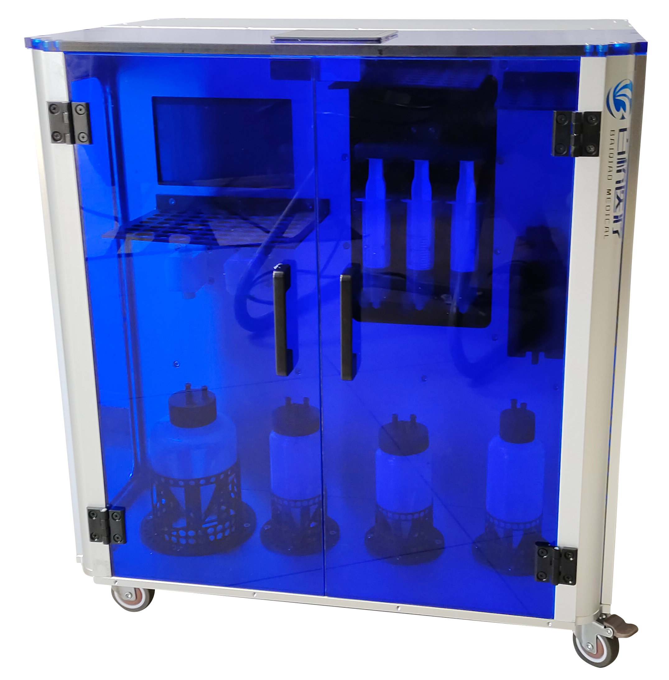
그림1 전자동 엑소좀추출·농축장비
Fig.1 Automatic Exosome Filtration Instrument
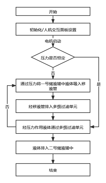
그림2 장비 작업 흐름도
Fig.2 Instrument work flow chart
1.2 장비 성능 평가
1.2.1 실험 재료
1.2.1.1 주요 실험 시약
지방줄기세포 배양 첨가제(UltraGROTM,AventaCell BioMedical, 미국);PBS(Gibco, 미국);0.25%트립신(Gibco, 미국); 소태아혈청(Gibco, 미국); 삼중항생제(Gibco,미국);α-MEM, 저당 및 고당DMEM(Gibco, 미국); 콜라게나아제(Collagenase NB4,Serva, 독일); BCA단백질 정량 키트(Beyotime, 중국).
1.2.1.2주요 실험 장비
일회용 멸균10 cm배양 접시(BD, 미국);100 kDa한외여과 튜브(Milipore, 미국);클린벤치(Suzhou Purification Equipment Co., Ltd., 중국); 마이크로플레이트 리더(Thermo Fisher Scientific, 미국);미량 전자저울(Longcheng Instrument Equipment Co., Ltd., 중국); 유세포분석기(Beckman,미국);온도 조절 탁상형 원심분리기(Thermo Fisher Scientific, 미국);혈구계산판(Shanghai Qiujing Biochemical Reagent Instrument Co., Ltd., 중국);온도 조절 셰이커(Taicang Huamei Biochemical Instrument Factory, 중국);항온 수조(Shanghai Hetian Scientific Instruments Co., Ltd., 중국).
1.2.2 실험 방법
1.2.2.1지방줄기세포 분리·배양
본 연구의 지방은 환자 복부 또는 허벅지 안쪽에서 지방흡입술 후 폐기된 조직입니다. 환자는 의료 연구용 기증에 동의하는 서면 동의서를 작성했고 연구계획은 상하이교통대학교 의과대학 부속 제9인민병원 윤리위원회의 승인을 받았습니다.
기증 지방을 콜라게나아제로 소화하고 원심분리하여 일차 지방줄기세포를 추출했습니다. 세포를 지방줄기세포 배양 첨가제가 든α-MEM배지의10cm배양 접시에 접종하여5%CO2,37℃에서 배양했습니다. 세포가 약80%-90%융합도에 도달하면EDTA함유0.25%트립신으로 세포를 소화하여 계대 배양합니다. 제3세대 지방줄기세포로 엑소좀을 추출합니다.
1.2.2.2지방줄기세포 유래 엑소좀 추출
수집한 제3세대 지방줄기세포 상등액을 엑소좀 추출 장비의 공급 탱크에 넣어 여과합니다. 얻은 여과액을100kDa함유milipore한외여과 튜브에 넣고 한외여과법으로 엑소좀을 추출합니다.3000g,15-20min,4℃에서 원심분리하여 한외여과 튜브 내관의 상등액이 거의 모두 내려갈 때까지 처리한 후PBS로 한 번 세척하고 내관에200-500μL엑소좀 함유PBS만 남을 때까지 원심분리합니다. 완료 후 엑소좀 함유PBS를 흡인하여4℃ 냉동고에 보관합니다.
1.2.2.3지방줄기세포 유래 엑소좀 동정
현재 분리법으로 매우 순수하고 단일한 엑소좀을 얻기는 어려우므로 장비 농축과 원심분리로 얻은 엑소좀을 동정해야 합니다. 본 연구는 국제세포외소포학회 기준에 따라 정확성을 보장하는 세 가지 방법을 사용했습니다.① 투과전자현미경(TEM)으로 전처리 엑소좀 형태 관찰[18];② 크기 기반 나노입자 추적 분석(NTA): 엑소좀 입자에 광선을 조사하고 산란광과 브라운 운동으로 각 입자의 경로를 기록합니다. 평균 속도와 확산계수를 구하고 수학적 계산으로 농도와 크기 분포를 산출합니다.[19];③ 웨스턴 블롯(Western Blot)으로 엑소좀 표면 특이 단백질 검출marker [20]。
1.2.3 주요 관찰 지표
①투과전자현미경지방줄기세포 유래 엑소좀 형태 관찰.②나노입자 추적 분석으로엑소좀 입자 크기와 농도 분석.③ 웨스턴 블롯으로 지방줄기세포 유래 엑소좀 발현 단백질 동정.
2 결과
2.1 지방줄기세포 형태
그림3(a)는 세척·정제된 지방이며, 그림3(b)은 도립현미경에서 관찰한 3세대 사람 지방줄기세포로 긴 방추형입니다.
2.2 엑소좀 투과전자현미경 동정 결과
그림4(a)와 같이,투과전자현미경에서 지방줄기세포 유래 엑소좀 크기는30nm-200nm사이였고 전형적인 원형 컵 모양이었습니다.
2.3 엑소좀 표지자 웨스턴 블롯 동정 결과
국제세포외소포학회 기준[21]에서는 소포를 엑소좀으로 정의하려면 엔도솜 또는 엑소좀 원형질막 단백질과 분비 관련 세포질 단백질을 포함하고, 비엑소좀 구조 또는 관련 광범위 세포질 단백질 하나를 배제해야 합니다. 그림4(b)와 같이 웨스턴 블롯에서 엑소좀 원형질막 단백질CD63、CD81발현과 분비 관련ESCRT복합체 단백질 중TSG101발현이 검출되었고 골지체 단백질GM130은 거의 발현되지 않아 분리 산물에 고순도 엑소좀이 포함되었음을 입증했습니다.
2.4 엑소좀 나노입자 추적 분석 결과
그림5와 같이 나노입자 추적 분석의 입자 크기 분포는30nm-200nm사이에 집중되었으며 농도 결과도 엑소좀 크기 특성에 부합했습니다.[22]。
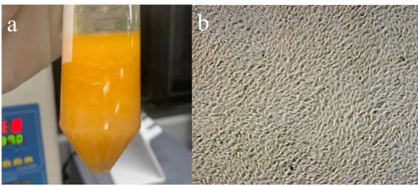
그림3 실험 재료. (a) 실험용 지방; (b) 사람 지방줄기세포 형태
Fig.3 Experimental materials.(a)Fat used in experiments;(b)Morphology of human adipose stem cells
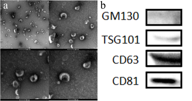
그림4 엑소좀 형태 및 특이 단백질 동정. (a) 투과전자현미경의 엑소좀 형태 특성; (b) 웨스턴 블롯으로 엑소좀 단백질 발현 검출
Fig.4 Exosome morphology and specific protein identification.(a)Morphological characteristics of exosomes displayed by transmission electron microscopy;(b)Western blotting to detect exosome protein expression
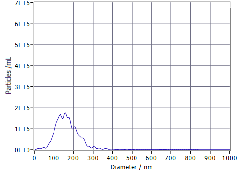
그림5 NTA 엑소좀 입자 크기 분포 검사
Fig.5 NTA detection of exosome particle size distribution
3 고찰
다단 여과 전자동 엑소좀 분리 장비의 성능 평가에는 자가 지방줄기세포를 원료로 사용했습니다. 자가 지방은 공급원이 풍부하고 쉽게 얻을 수 있으며 면역 거부반응이 없어 적용이 편리하다는 임상적 의미가 있습니다. 또한전자동 다단 여과 엑소좀 분리 장비는 비접촉 설계와 지능형 폐루프 제어를 적용해 인건비를 절감하고 고순도·무오염 엑소좀을 추출하여 임상 기준을 충족합니다.평가 결과, 지방줄기세포 유래 엑소좀을 농축·원심분리하고 세 가지 표준법으로 동정한 결과 추출 미립자가 엑소좀이며 오염도가 정상 범위였습니다. 장비로 엑소좀 함유 상등액을 여과·농축한 뒤 분리하면 완전한 엑소좀을 얻을 수 있고 처리 시간이 기존 방법보다 훨씬 짧아 임상 적용 요건을 충족함을 입증했습니다.
이 장비는 서로 다른 엑소좀 아형의 입자 크기(30-150nm)별 정밀 세분 추출을 실현하여 아형 구조와 기능의 관계 연구에 유용한 도구를 제공하고 임상 치료에 신속하고 효율적인 방법을 제공합니다. 다만 일부 엑소좀이 필터 막에 부착해 산물·활성이 손실되는 문제가 있습니다. 향후 접선류 등 새로운 여과법으로 장비를 개선하여 농축액 낭비를 줄이고 회수량을 높일 예정입니다.
4 결론
전자동 다단 여과 장비는 엑소좀을 빠르고 효율적으로 분리하여 임상 적용 요건을 충족하고 엑소좀 기반 질환 치료를 지원합니다.
참고문헌
[1] Raposo G,Stoorvogel W. Extracellular vesicles: exosomes, microvesicles, and friends.[J]. The Journal of cell biology,2013,200(4).
[2] Lee Y,El A S,Wood MJA. Exosomes and microvesicles: extracellular vesicles for genetic information transfer and gene therapy.[J]. Human molecular genetics,2012,21(R1).
[3] Marqués-García F,Isidoro-García M. Protocols for Exosome Isolation and RNA Profiling.[J]. Methods in molecular biology (Clifton, N.J.),2016,1434.
[4] Li P,Kaslan M,Lee SH,et al. Progress in exosome isolation tech-niques[J]. Theranostics,2017,7( 3) : 789-804.
[5] Clark DJ,Fondrie WE,Liao Z,et al. Redefining the breast cancer exosome proteome by tandem mass tag quantitative proteomics and m*riate cluster analysis[J]. Anal Chem,2015,87(20) : 10462-10469.
[6] Yu B,Zhang X,Li X. Exosomes derived from mesenchymal stem cell[J]. Int J Mol Sci,2014,15(3) : 4142-4157.
[7] Ye Dongman,Yu Tao. 종양 엑소좀의 추출 및 동정[J]. 현대 종양의학,2018,26(20),3341-3343.
[8] Chan BD,Wong WY,Lee MM,et al. Exosomes in inflammation and inflammatory disease[J]. Proteomics,2019,13: e1800149.
[9] MATHIEU M, MARTIN-JAULAR L, LAVIEU G, et al. Specificities of secretion and uptake of exosomes and other extracellular vesicles for cell-to-cell communication [J]. Nat Cell Biol, 2019, 21(1): 9-17.
[10] SHU S, YANG Y, ALLEN C L, et al. Purity and yield of melanoma exosomes are dependent on isolation method [J]. J Extracell Vesicles, 2020, 9(1): 1692401.
[11] Shi MM, Yang QY, Monsel A, et al. Preclinical efficacy and clinical safety of clinical-grade nebulized allogenic adipose mesenchymal stromal cells-derived extracellular vesicles. J Extracell Vesicles. 2021;10(10):e12134.
[12] Zhao L, Gu C, Gan Y, Shao L, Chen H, Zhu H. Exosome-mediated siRNA delivery to suppress postoperative breast cancer metastasis. J Control Release. 2020;318:1-15.
[13] Jiang WJ, Xu CT, Du CL, et al. Tubular epithelial cell-to-macrophage communication forms a negative feedback loop via extracellular vesicle transfer to promote renal inflammation and apoptosis in diabetic nephropathy. Theranostics. 2022;12(1):324-339.
[14] Jiang L, Fei H, Jin X, et al. Extracellular Vesicle-Mediated Secretion of HLA-E by Trophoblasts Maintains Pregnancy by Regulating the Metabolism of Decidual NK Cells. Int J Biol Sci. 2021;17(15):4377-4395.
[15] WEN S W, LIMA L G, LOBB R J, et al. Breast cancer-derived exosomes reflect the cell-of-origin phenotype [J]. Proteomics, 2019, 19(8): e1800180.
[16] Yang Dongbin,Zhang Weihong,Zhang Huanyun,Zhang Fengqiu,Chen Lanmei,Ma Lixia,Larcher Leon M,Chen Suxiang,Liu Nan,Zhao Qingxia,Tran Phuong H L,Chen Changying,Veedu Rakesh N,Wang Tao. Progress, opportunity, and perspective on exosome isolation - efforts for efficient exosome-based theranostics.[J]. Theranostics,2020,10(8).
[17] Bai Jun,Ren Jun,Wang Jun,Zhang Hongmei,Yang Jingyue,Zhang Liwang.원심여과법을 이용한 엑소좀 추출 실험 연구[J].제4군의대학교 학보,2004(10):890-892.
[18] Lässer C,Eldh M,Lötvall J. Isolation and characterization of RNA-containing exosomes.[J]. Journal of visualized experiments : JoVE,2012(59).
[19] Xiao-Xia Yang,Chao Sun,Lei Wang,Xiu-Li Guo. New insight into isolation, identification techniques and medical applications of exosomes[J]. Journal of Controlled Release,2019,308(C).
[20] Théry C,Amigorena S,Raposo G,Clayton A. Isolation and characterization of exosomes from cell culture supernatants and biological fluids.[J].Current protocols in cell biology,2006,Chapter 3(1).
[21] Théry C, Witwer KW, Aikawa E, et al. Minimal information for studies of extracellular vesicles 2018 (MISEV2018): a position statement of the International Society for Extracellular Vesicles and update of the MISEV2014 guidelines. J Extracell Vesicles. 2018;7(1):1535750. Published 2018 Nov 23. doi:10.1080/20013078.2018.1535750
[22] Wang Shuai,Li Chaoran,Zhang Qianru,Huang Guilin.사람 소변 유래 줄기세포 엑소좀의 추출 및 동정[J].중국 조직공학 연구,2022,26(13):2056-2061.