
 You Are Here:
Inside Vision Technology: A Step-by-Step Guide to Precise Operation of a Quantitative Corneal Transparency Measurement Instrument
 Updated: 2025-12-24
 Views: 282
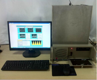
Quantitative corneal transparency meteris ophthalmic testing equipment [the source modifier is unclear] for objective, precise assessment of corneal optical clarity. It is used mainly in research, clinical diagnosis, and monitoring of corneal disease or postoperative recovery. As the transparent tissue at the front of the eye, corneal clarity directly affects visual quality; inflammation, edema, scarring, dystrophy, or surgical trauma can reduce transparency and impair vision. Traditional examinations often rely on subjective slit-lamp microscopy and lack quantitative standards, whereas a quantitative corneal transparency meter uses optical principles for non-invasive, repeatable, digital precision assessment.
Quantitative corneal transparency meteroperating method:
I. Equipment preparation
Check equipment status: ensure that power is connected and the equipment is switched on. Check the display and control panel and confirm that all display and control functions operate normally.
Prepare accessories: according to measurement needs, prepare the measurement head, eye mask, and other necessary accessories and ensure that they are clean.
Prepare the environment: avoid direct intense light and electromagnetic interference and maintain stable ambient temperature and humidity to ensure accurate results.
II. Patient preparation
Adjust the patient's position: seat the patient in front of the equipment and adjust seat height and position so the eye is level with the measurement probe.
Stabilize the head: use the equipment's head support to keep the patient's head stable throughout measurement.
Fixation target: instruct the patient to look through the eyepiece at the target, generally a small red light or image, to keep the eye stable for measurement.
III. Calibration (if required)
Warm-up: perform the required warm-up according to the instrument manual so the equipment reaches a stable operating state.
Calibration: use a reference sample or follow the device prompts. For example, with no sample present, start acquisition and check whether the transparency displayed on the front panel is 1, or approximately 1. If not, finely adjust the beamsplitter holder angle until the expected transparency appears.
IV. Measurement
Place the sample (if applicable): when measuring a specific sample such as decellularized porcine corneal matrix, open the test-chamber lid, place the sample on the stage with forceps, and close the lid to prevent ambient light from affecting the reading.
Enter parameters: enter the sample thickness, if applicable, in the medium-thickness field so the equipment can calculate accurately.
Start measurement: run the program and click the acquisition button. The equipment automatically directs a standardized light source at the cornea or sample and records the intensity transmitted through it.
Maintain stability: keep the instrument stable during measurement so vibration or movement does not affect results. Instruct the patient to keep the eye still and avoid blinking.
V. Data reading and recording
Read data: after measurement, the display shows results including the quantitative corneal transparency value.
Record data: accurately read and record transparency, brightness, and other parameters, together with the test time and patient information.
Repeat measurement (if needed): adjust conditions or repeat the measurement as necessary to obtain more accurate and reliable results.
VI. Cleaning and maintenance
Clean the equipment: after testing, clean the test platform, photosensitive element, and other components for the next use.
Regular maintenance: follow the manual's requirements, including checks for damaged components and loose fastening screws, to ensure stable long-term operation.